Atopic dermatitis in dogs is one of the most complex and common dermatological conditions encountered in daily practice. It is a chronic, genetically based inflammatory condition, characterised by pruritus and skin lesions secondary to the immune response to environmental allergens and disruption of the epidermal barrier.
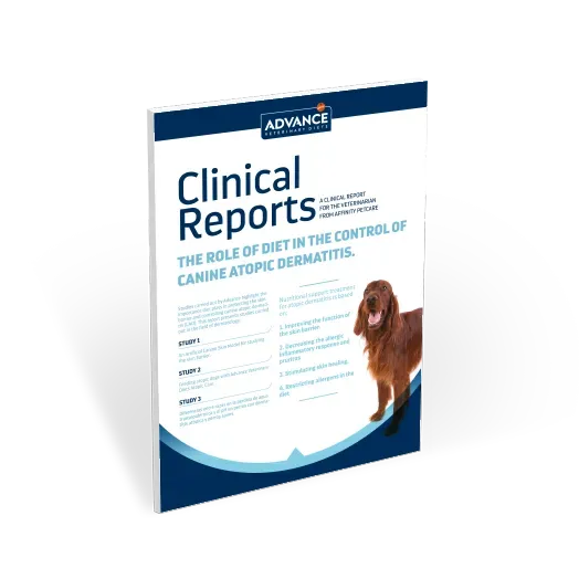
A multimodal approach to canine atopic dermatitis
Treatment of an acute flare-up
The primary objective is to halt the inflammatory cascade and provide immediate relief from the dog’s pruritus:
- Specific therapy: administration of oclacitinib, prednisone/prednisolone and management of secondary infections (bacterial or fungal).
- Supportive therapy: initiation of a tailored diet (with antigenic restriction and essential fatty acids) alongside frequent bathing with a specific shampoo.
Long-term management and maintenance
The aim is to prevent relapses, restore the epidermal barrier and reduce dependence on systemic drugs.
- Specific therapy: use of ciclosporin or allergen-specific immunotherapy.
- Supportive therapy: maintenance with a tailored diet, a schedule of weekly or fortnightly baths, and strict control of ectoparasites and endoparasites.
The importance of diet for dogs with atopic dermatitis
In canine atopic dermatitis, supportive treatment is key to minimising the long-term use of corticosteroids.
Advanced dietary management
A diet formulated for dogs with atopic dermatitis, containing essential fatty acids and functional nutrients (such as aloe vera), acts on three levels:
- Restoration of the skin barrier: it promotes the synthesis of lipids (ceramides, cholesterol and free fatty acids) in the stratum corneum.
- Corticosteroid-sparing effect: reduces the total cumulative dose of cortisone by up to 40% during flare-ups.
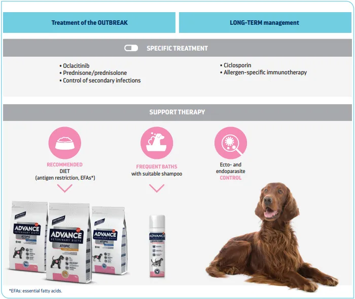
- Symptom control: it significantly reduces CADESI-03 scores (lesion severity) and pruritus visual analogue scale (PVAS) scores.
Tailored topical therapy
Regular use of a shampoo for atopic dermatitis in dogs helps to remove allergens from the skin’s surface, rehydrate the skin and stabilise the epidermal pH. It is important to remember that parameters such as skin pH or transepidermal water loss (TEWL) vary considerably depending on the breed (with particularly pronounced alterations in breeds such as the French Bulldog compared with the Cocker Spaniel).
What is the best food for dogs with atopic dermatitis? Advance Veterinary Diets Atopic
Affinity’s scientific team has focused its research on developing a well-tolerated diet that helps manage canine atopic dermatitis by restoring skin integrity and reducing symptoms.
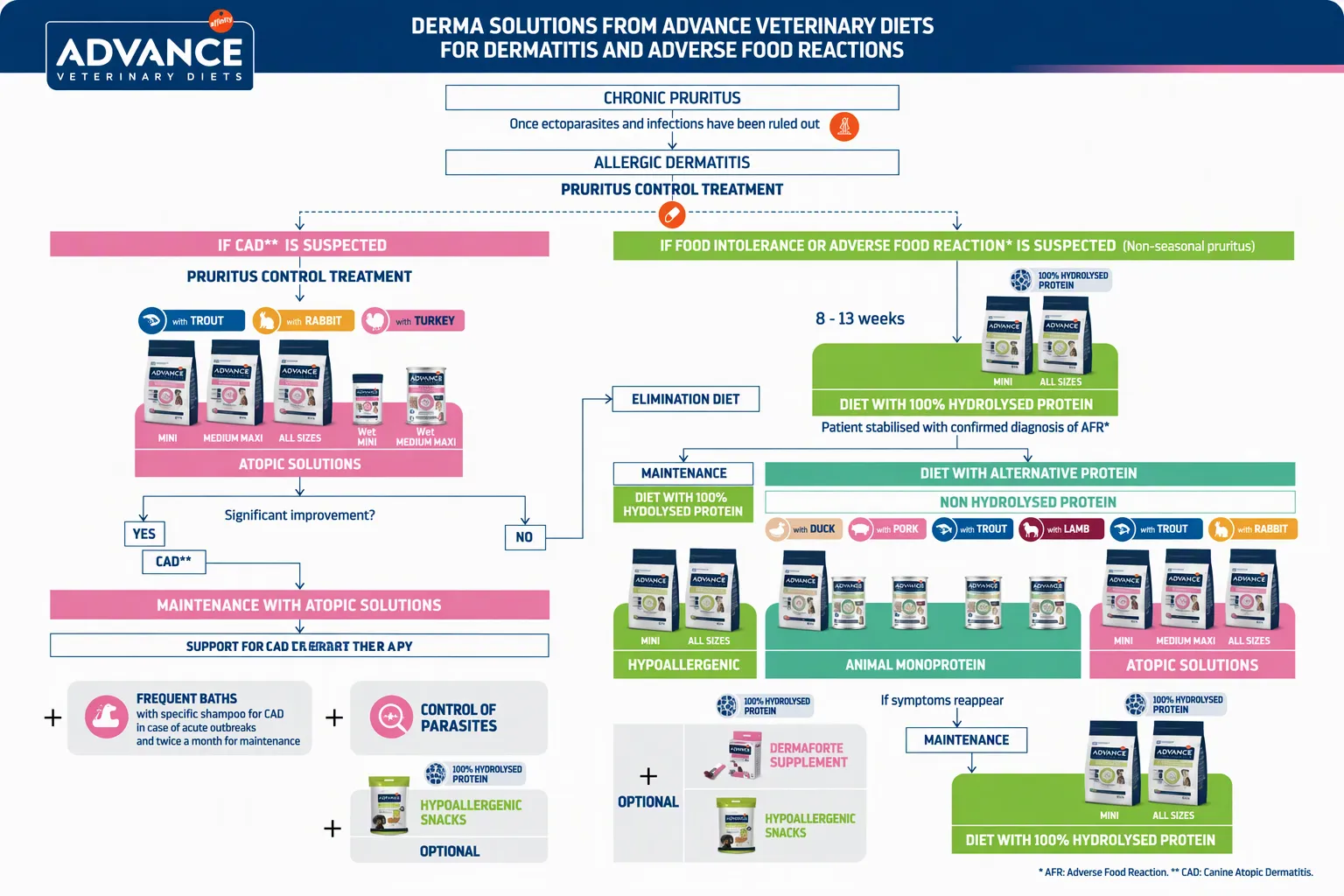
Choose the most suitable option from the Advance Atopic range according to the patient’s profile, size and any suspected concurrent food intolerances:
- Atopic Mini Trout: adapted to the nutritional needs of small-breed dogs, with appropriately sized kibble.
- Atopic Medium-Maxi Trout: specifically formulated to meet the needs of medium- and large-breed dogs.
- Atopic Rabbit: an alternative for patients who may benefit from rabbit as a novel protein.
- Atopic Wet: a highly palatable wet food, perfect for combining with dry food.
- DermaForte supplement: with extra essential fatty acids, ideal for complementing the Advance Veterinary Diets Atopic product range.
Combine the diet with the topical and hygiene range to maximise the recovery of the skin barrier. To find the most suitable option, you can use this decision tree:
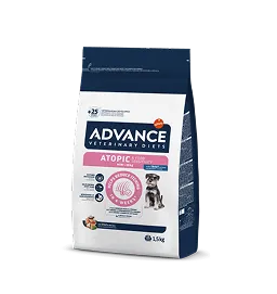
Advance Veterinary Diets Atopic
Low-allergen food that protects the skin barrier, promotes healing, and helps reduce itching and the inflammatory response.
Frequently Asked Questions
How do I explain to the owner the need to change the dog’s diet if the dog is already taking corticosteroids or oclacitinib?
You should explain that the medication puts out the ‘fire’ (the acute itching), but the special diet provides the building blocks (essential fatty acids) to repair the skin barrier.
Can Atopic dry food be mixed with home-cooked food or commercial treats during treatment?
This is not recommended, as introducing additional, unselected protein sources or an unbalanced fat intake invalidates the antigenic restriction and alters the optimal ratio of essential fatty acids that protect the skin barrier. If the owner needs to give treats or wet food, you can recommend other products from the Atopic range, formulated for animals requiring an extra supply of essential fatty acids. Always ensure that combining the food with other sources of animal protein does not pose a problem for the animal.
References
1. Marsella R & Samuelson D. Unraveling the skin barrier: a new paradigm for atopic dermatitis and house dust mites. Veterinary Dermatology 20, 533-540, 2009.
2. Oyoshi MK, He R, Kumar L, Yoon J, Geha RS. Cellular and molecular mechanisms in atopic dermatitis. Advances in Immunology 102, 135-226, 2009
3. Olivry T, Deboer DJ, Favrot C, Jackson HA, Mueller RS, Nuttall T, Pr.laud P, International Task Force on Canine Atopic Dermatitis. Treatment of canine atopic dermatitis: 2010 clinical practice guidelines from the International Task Force on Canine Atopic Dermatitis. Veterinary Dermatology 21, 233-248, 2010.
4. Serra M., Braz.s P., Puigdemont A., Fondevila D., Romano v., Torre C., Ferrer Ll. 2007. Development and characterization of a canine skin equivalent. Experimental Dermatology 16, 135- 142, 2007.
5. Olivry T (Ed.). The American College of Veterinary Dermatology Task Force on Canine Atopic Dermatitis. Veterinary Immunology Immunopathology Special Issue, Vol 81, 2001.
6.Hillier A, Griffin CE. The ACVD task force
on canine atopic dermatitis (X): is there a relationship between canine atopic dermatitis and cutaneous
adverse food reactions?. Veterinary Immunology Immunopathology 81, 227-231, 2001.
7. Cornegliani L et al; Transepidermal water loss in healthy and atopic dogs, treated and untreated: a comparative preliminary study. Vet Dermatology 2012 Feb;23(1):41-4
8. Shimida K et al; Increased transepidermal water loss and decreased ceramide content in lesional and non-lesional skin of dogs with atopic dermatitis. Vet Dermatology 2009 Oct;20(5-6):541-6.
9. Rippke F et al. Stratum corneum pH in atopic dermatitis: impact on skin barrier function and colonization with Staphylococcus Aureus. Am J Clin Dermatol. 2004;5(4):217- 23.