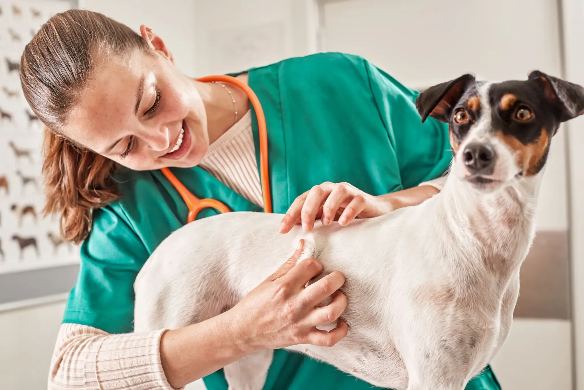
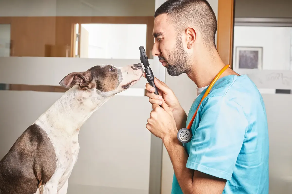
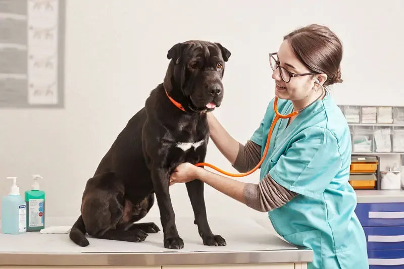
Introduction
Mites are small parasites (<1 mm long) with three pairs of legs in the larval stage and four in the nymph and adult stages. They belong to the phylum Arthropoda , class Arachnida and subclass Acari . In dogs, they are responsible for several diseases , some of which also affect humans .
Therefore, vets need to be familiar with the diagnosis and treatment of mite-related diseases in dogs.1
Main diseases caused by mites in dogs
Some of the diseases caused by mites in dogs include sarcoptic mange, canine demodicosis, cheyletiellosis, otodectic mange, trombiculosis and nasal mite lesions, which are discussed briefly below.
- Sarcoptic mange , caused by Sarcoptes scabiei var. canis , is a transmissible, nonseasonal and extremely pruritic disease with zoonotic potential . It mainly affects young dogs that frequently visit parks and mix with peers. Up to 70% of patients have lesions on the face or pinnae . Other commonly affected areas of the body include the ventral abdomen, thorax, elbows, hocks and digits.2,3
- Demodecic mange is caused by excessive proliferation of the mite Demodex canis , although D. injai and D. cornei have been reported.In puppies , it is believed that a temporary deficiency in cell-mediated immunity leads to the disease’s onset, whereas in adult dogs , it is associated with “pharmacological” immunosuppression or the presence of other diseases. Demodicosis in dogs can be localised , with spontaneous remission in most cases without any specific treatment; or generalised , with a much severer clinical picture , which in extreme cases could result in the death/euthanasia of the animal. Initial signs include inflammatory or non-inflammatory hypotrichosis/alopecia with the formation of comedones and flaking . Hyperpigmentation may also be observed. Pruritus, when present, is usually associated with the presence of short forms of D. canis or a secondary bacterial infection.2-4
- Cheyletiellosis , or walking dandruff, is a nonsuppurative pruritic dermatitis which is caused by Cheyletiella yasguri in dogs and can also affect humans. It is an extremely contagious disease , especially among puppies living in kennels or shelters.2,3
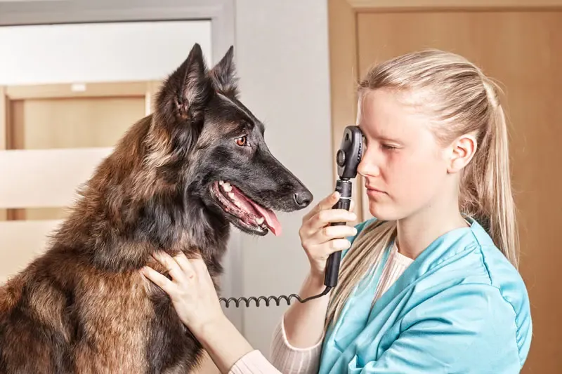
- The causative agent of otodectic mange in dogs is Otodectes cynotis (ear mite) . Ear mites cause itching of varying intensity. Otoscopic examination often reveals an ear canal filled with earwax, remnants of blood and parasite waste, giving the discharge the characteristic appearance of “coffee grounds” . O. cynotis can be isolated from other areas of the body, where it may not cause clinical signs or pruritic dermatitis. In humans, it can cause a transient papular dermatitis and in rare cases develop into an otic parasite.2,3
- Trombiculosis in dogs is caused by Neotrombicula ( Trombicula ) autumnalis and Straelensia cynotis , also known as harvest mites or red mites .
- N. autumnalis causes very pruritic lesions, usually affecting the head, ears, limbs and interdigital spaces .
- Straelensiosis usually affects rural dogs that live outdoors in late summer or early autumn. It has been reported mainly in Spain, Portugal, France and Israel. Affected animals usually have a characteristic light to deep red papular/nodular dermatitis that evolves into dark papular crusts and mainly affects the head and back, although sometimes it can be generalised .2,3,5
- Pneumonyssoides caninum , ( nasal mites ) mainly infects large dogs, over 3 years old . It normally inhabits the nasal cavity and sinuses, but it does not always produce any clinical signs. When present, the clinical picture is characterised by sneezing and reverse sneezing, serous/catarrhal rhinosinusitis , facial pruritus and aerophagia, which may predispose the development of gastric torsion.3,6
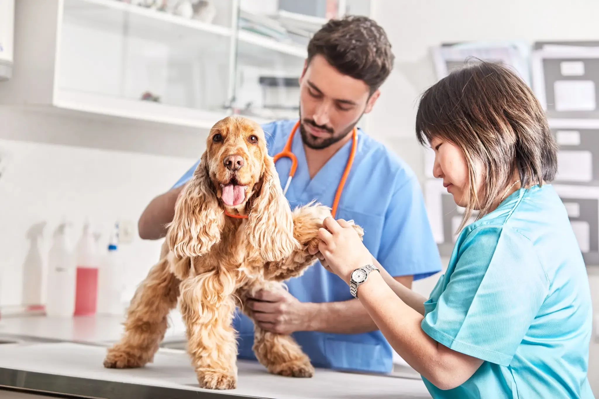
Diagnosis of mite-related diseases in dogs
In most of these infestations, the diagnosis is based on the identification of the mite in samples of superficial or deep skin scrapings (depending on the suspected mite being sought), the tape method or by performing a trichogram .
P. caninum can usually be detected during a rhinoscopy or identified in samples from nasal lavages . If ingested while licking and excreted in faeces, some of these mites can also be identified by stool analysis . Intradermal and serological tests are also available in the case of S. scabiei .Finally, in some mites that are difficult to identify, such as S cynotis , the definitive diagnosis can be reached through a skin biopsy .2,3,5,6
Treatment
A detailed description of the treatment of each of these diseases is beyond the scope of this article and the reader is advised to consult the referenced literature.
Nevertheless, it is important to know that different treatment options are available of varying efficacy depending on the disease. The most commonly used drugs include amitraz (demodicosis), fipronil (cheyletiellosis, otodectic mange, trombiculosis due to N. autumnalis ), macrocyclic lactones (sarcoptic mange, otodectic mange, demodicosis, nasal mites, cheyletiellosis), pyrethroids (trombiculosis due to N. autumnalis ) and, more recently, isoxazolines (sarcoptic mange, otodectic mange, demodicosis, trombiculosis), which have become the best weapon available to the vets.
In addition to acaricidal treatment, in case of very intense pruritus and/or secondary infections , it is a good idea to administer drugs that help control itching , as well as the appropriate antibiotic therapy .2,3,5
Conclusions
Most vets will probably have to treat a variety of diseases caused by mites over the course of their career. Microscopic examination of skin scrapings or exudates is often very helpful in the diagnosis and should therefore be a standard test whenever a patient has a compatible clinical picture. Regarding treatment, and although there are different therapeutic options, isoxazolines have truly revolutionised the management of many of these diseases, thanks to their high efficacy and ease of administration.