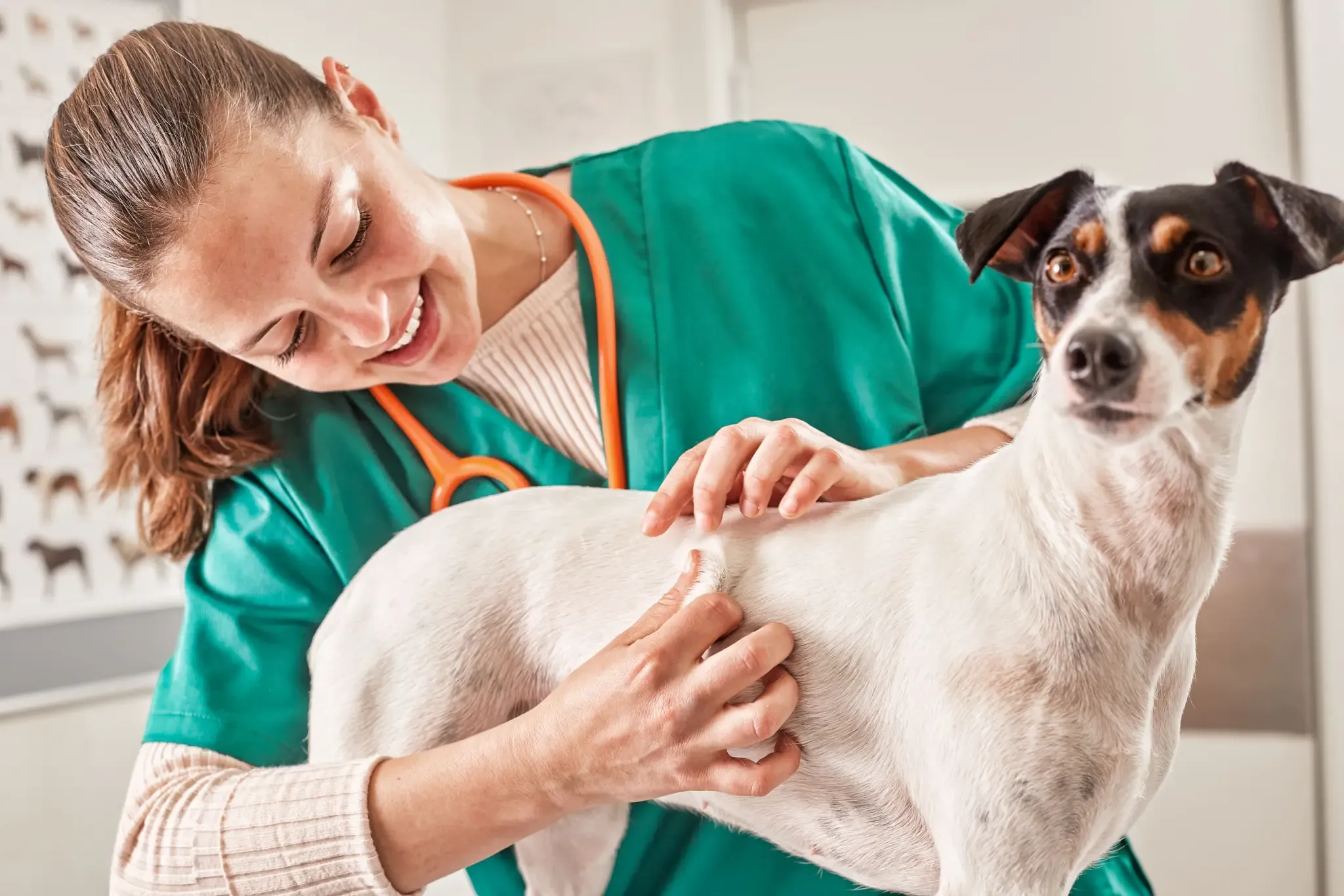
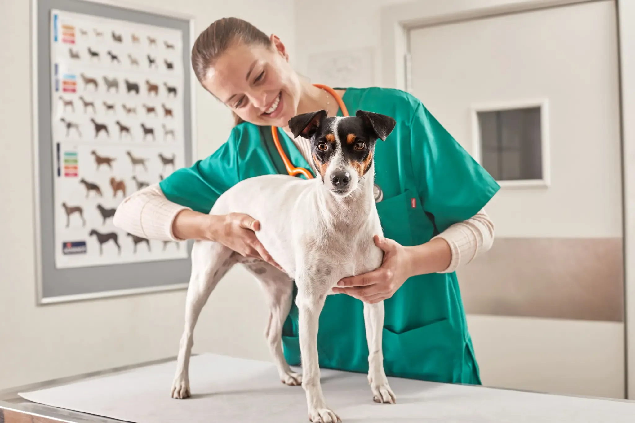
What is hypopigmentation or vitiligo in dogs?
Vitiligo is characterised by the complete absence of melanocytes, with a normal dermis and epidermis. It has an unknown aetiology and courses asymptomatically. Some of the proposed aetiologies include an autoimmune, toxic or nervous origin triggered by a virus (for more information on dermatology see this Affinity Advance Clinical Report).
- Differential diagnosis : It is important to differentiate hypopigmentation in dogs from other conditions that cause seasonal nasal depigmentation , which is a temporary reduction (though not complete absence) of melanocytes leading to seasonal depigmentation and repigmentation.
Main causes of hypopigmentation in dogs
Hereditary causes
- Albinism: total absence of pigment.
- Piebaldism: limited areas with no melanocytes.
- Klein–Waardenburg syndrome: affected animals have areas of skin and coat without melanocytes, and blue eyes or heterochromia accompanied by deafness. Reported in cats, Bull Terrier, Sealyham Terrier, Collie and Dalmatian.
- Canine cyclic haematopoiesis: a fatal autosomal recessive disease. Collies develop a grey coat, light-coloured nose and cyclical episodes of neutropaenia every 12–14 days that cause sepsis and amyloidosis.
- Chediak–Higashi syndrome: a rare autosomal recessive disease in Persian cats. It is characterised by partial albinism of the eyes and skin associated with abnormal granulocytes and platelets, causing bleeding, recurrent infections and death in the first months of life.
- Greying: age-related decline in melanocyte replication.
- Vitiligo: white macules on the nose, ears, oral mucosa and face. It is caused by antimelanocyte antibodies. Mainly observed in Siamese cats, Belgian Tervurens, German Shepherds, Collies, Rottweilers, Doberman Pinschers and Giant Schnauzers.
- Nasal hypopigmentation: seasonal clearance of the nasal planum during winter, primarily seen in the Siberian Husky, Golden Retriever, Labrador and Bernese Mountain Dog.
Acquired causes
- Postinflammatory: the leading cause in this subgroup is discoid lupus erythematosus, which causes nasal depigmentation. Other causes include pemphigus, systemic lupus erythematosus, uveodermatologic syndrome, bullous pemphigoid, mucocutaneous pyoderma, toxic rash and contact dermatitis. Some infectious causes are leishmaniasis, blastomycosis, sporotrichosis and bacterial folliculitis.
- Toxic: caused by drugs such as ketoconazole, procainamide and vitamin E.
- Nutritional/metabolic: zinc, pyridoxine, pantothenic acid and lysine deficiencies. These make the coat turn greyish. Dark hairs can become reddish in cases of copper deficiency, hypothyroidism, hyperadrenocorticism, hyperoestrogenaemia and hyperprogesteronaemia or exposure to chlorine or ultraviolet light.
- Neoplastic: nasal depigmentation, leukoderma and occasionally cutaneous T-cell lymphoma, basal cell tumours, mammary adenocarcinoma and gastric carcinomas.
- Idiopathic: patchy hypopigmentation reported in breeds such as Labrador Retriever and Newfoundland. Siamese cats may also present periocular leukotrichia in association with upper respiratory tract infection, pregnancy, malnutrition or systemic disease.
Is there a treatment for vitiligo?
Hypopigmentation or vitiligo in dogs is a purely aesthetic condition that does not cause any harm to the animal and has no specific treatment , although cases of spontaneous repigmentation have been described.