The most frequent clinical signs of conjunctivitis in dogs include:
- Squinting or constant blinking .
- Excessive tearing .
- Redness .
- Swelling around the eyes.
- Discharge from the eyes (watery, mucoid or mucopurulent).
Conjunctivitis can affect one or both eyes. Leading causes of conjunctivitis are:
- Viral infections.
- Immune disorders: allergic conjunctivitis (atopy or seasonal allergies), plasma cell conjunctivitis and pemphigus.
- Eyelid and conjunctival tumours .
- Breed-related conditions (such as nodular episcleritis ).
- Tear film deficiency.
- Eyelid abnormalities: entropion or ectropion .
- Eyelash disorders: distichiasis and ectopic cilia .
- Nasolacrimal duct obstruction.
- Trauma or irritation due to foreign bodies or environmental contaminants.
- Other eye disorders: ulcerative keratitis , anterior uveitis and glaucoma .
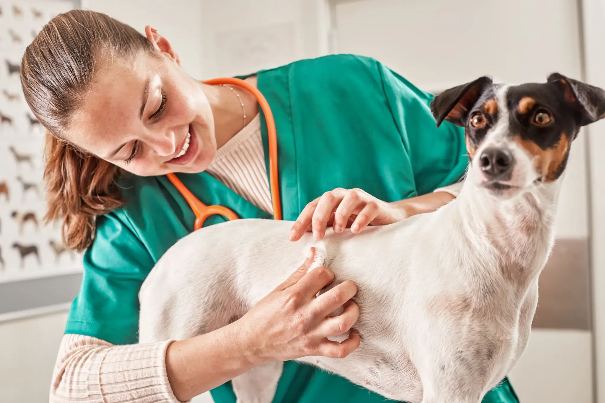
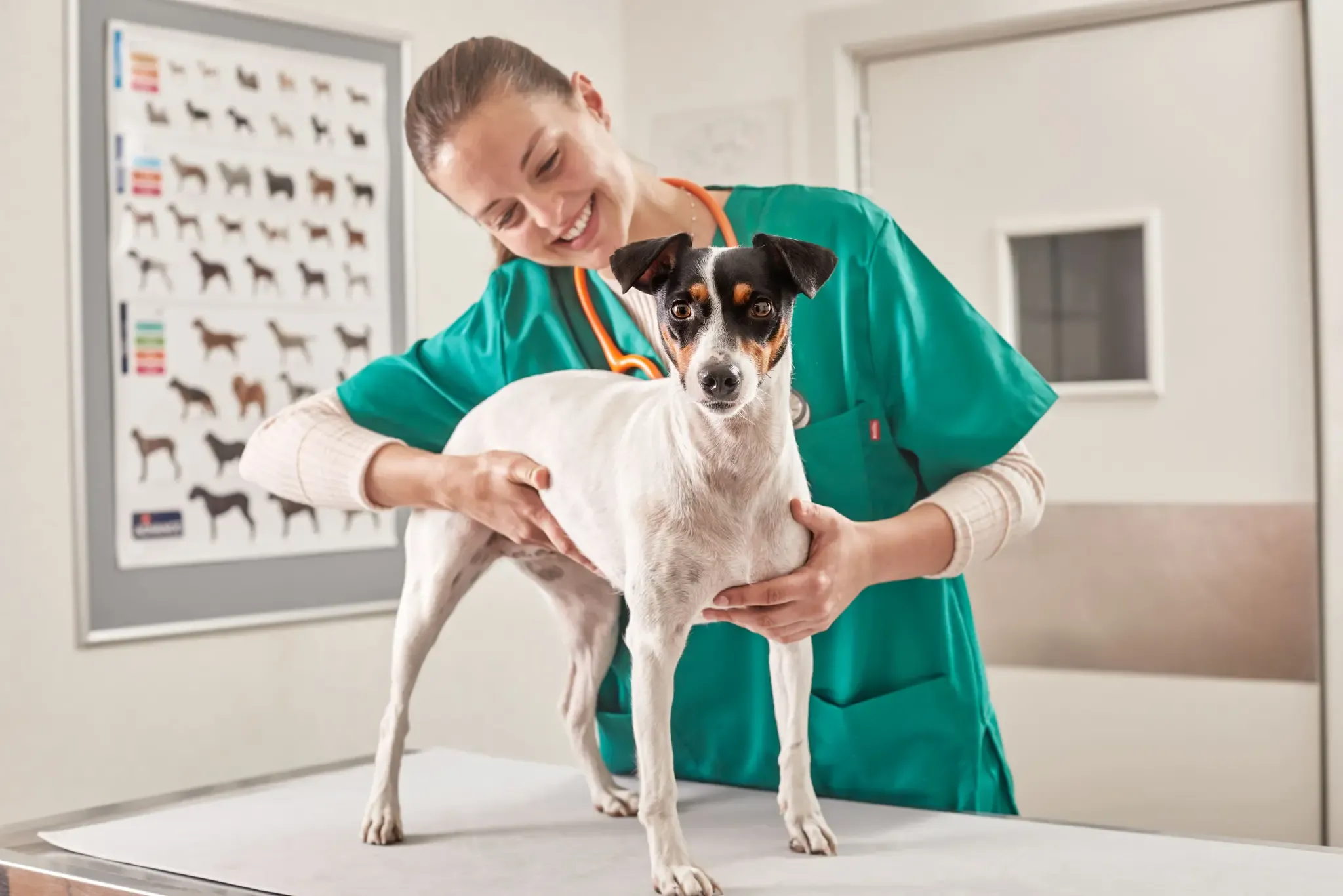
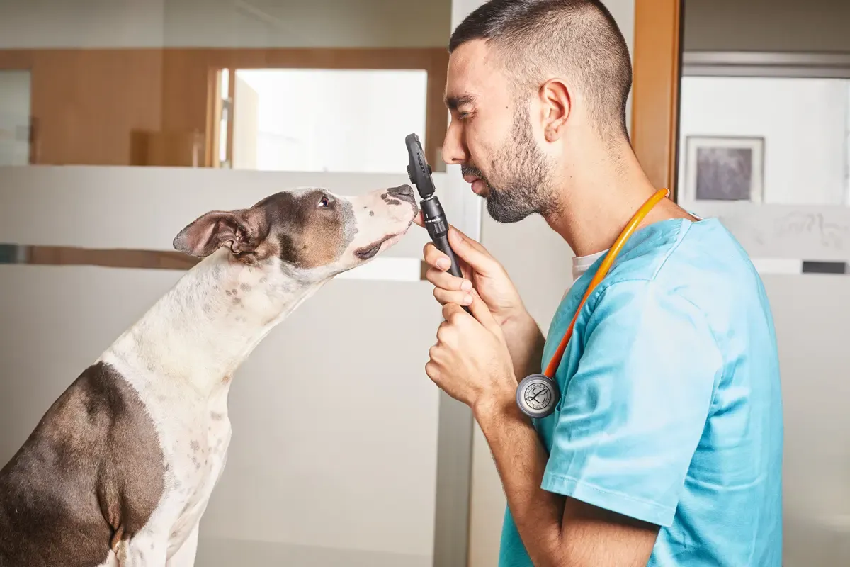
Conjunctivitis in dogs: diagnosis
Identifying the cause of conjunctival inflammation in dogs is key to its proper treatment . The vet needs to determine if the conjunctivitis is a primary or secondary problem, if there is any damage or concurrent eye disease, if the sclera is affected and if the condition is allergic, viral, or bacterial.
A detailed and complete ophthalmic examination is necessary to differentiate between these conditions. The most popular and clinically useful laboratory methods for assessing conjunctival specimens are microscopic study of cytology samples , culture and susceptibility testing , live virus isolation, polymerase chain reaction, antigen testing , and histopathological study for surgical biopsy .
This article focuses on corneal and conjunctival cytology as a diagnostic aid.
Corneal and conjunctival cytology as an aid in the diagnosis of conjunctivitis in dogs
A cytology study helps reach a quick and easy disease diagnosis. However, it should be complemented by other diagnostic laboratory techniques , such as culture and germ identification.
A local anaesthetic should not be used to collect the sample because they contain bacteriostatic or antiseptic agents that distort the results. First, the eyelids should be everted manually to avoid contamination. The back of a scalpel blade, sterile swab or cytology brush can be used for this manoeuvre. The chosen instrument is used to gently rub the conjunctival fornix between the third eyelid and the cornea or over the cornea.
After collecting the sample, smear it across a glass slide and leave it to air dry. The dried sample is then fixed with 5% methanol for about 5 minutes before Giemsa staining .
The next step is a microscope study with immersion objectives (1000x magnification). Inspect the area noting the most visible cells or structures.
The conjunctival scraping from a healthy dog should only contain epithelial cells and mucus. Signs of foreign organisms and/or inflammatory cells are indicative of disease . The most common inflammatory cells are lymphocytes, monocytes, plasma cells and neutrophils.
Lymphocytes and monocytes predominate in viral conjunctivitis , neutrophils in bacterial conjunctivitis, neutrophils with observation of basophilic intracytoplasmic inclusions in chlamydial conjunctivitis (cats), and eosinophils, basophils and neutrophils predominate in allergic conjunctivitis.
Conjunctivitis in dogs: treatment
Treatment can include topical and oral or systemic medications . Topical ointments and solutions of gentamicin, tobramycin, chloramphenicol, oxytetracycline, ciprofloxacin or triple antibiotic eye ointment are commonly prescribed. Sometimes medications containing anti-inflammatory agents , such as ophthalmic prednisolone or dexamethasone, are also administered. Dogs with eyelid or eyelash abnormalities require surgical correction.