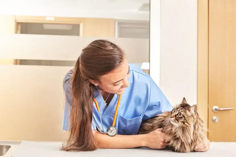
The sterilisation or castration of cats includes a wide variety of techniques intended to both alters the animal’s ability to reproduce and to prevent unwanted behaviours.1
So, in females we are referring to both ovariohysterectomies (OVH) and oophorectomies. Both the ovaries and the uterus are removed during an OVH, while only the ovaries are removed in an oophorectomy. The two procedures require different incisions; oophorectomies involve smaller incisions and cause lesser complications. The recommended age for castrating cats is 5–7 months.1
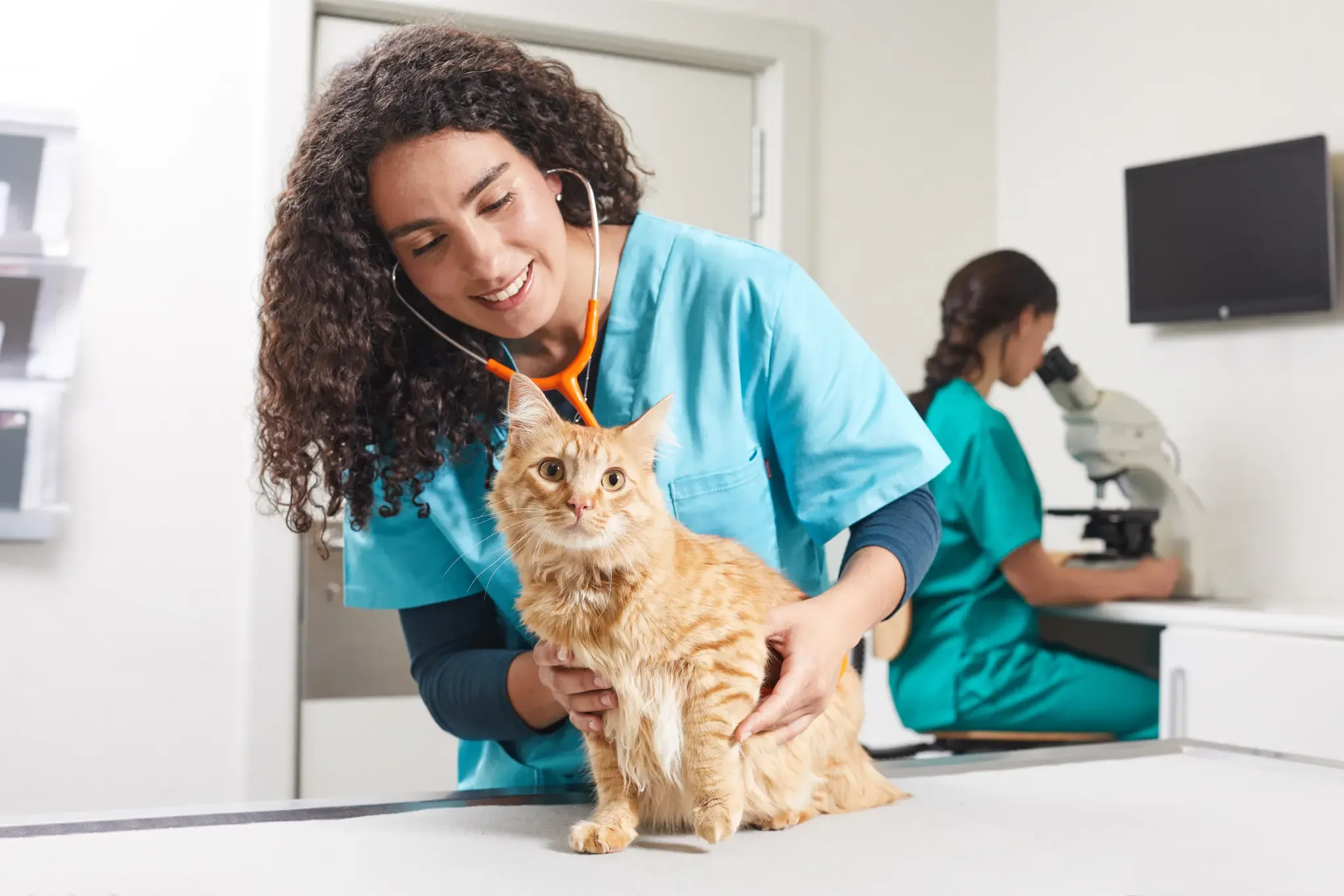
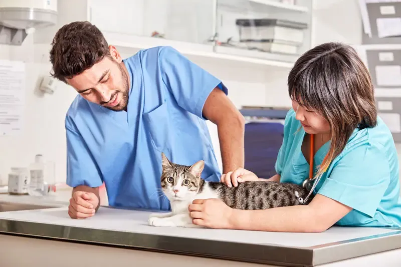
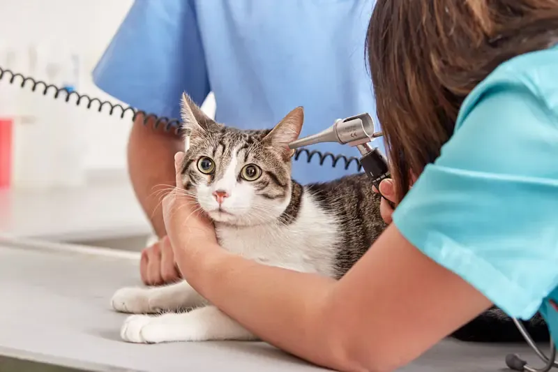
Preoperative considerations
As these interventions are also performed for reasons other than reproductive control, the preoperative period is the ideal time to conduct an appropriate study of the reproductive system based on the anamnesis, clinical signs, physical examination, diagnostic imaging, endoscopy, cytology, microbiology, hormone assays, haematology, blood chemistry profile, urinalysis and/or other laboratory tests.1
This study is essential to help mitigate possible future complications. Some potential complications, such as electrolyte or acid–base imbalances, are associated with the anaesthesia, which will be general in the case of nonessential surgeries. Which is why we have anaesthetic protocols in place specifically to avoid complications.1
Castration
A correct procedure also entails appropriate preoperative care :1
- Patients must fast for 12–18 hours (4–8 hours for paediatric animals) prior to surgery.
- In addition, the urinary bladder must be emptied manually if the patient has not done so before induction.
There are variants on the standard OVH technique, including flank and laparoscopic approaches and the use of staples, ultrasonic scalpels, vessel sealing systems, transfixing ligatures or Miller’s knots. The technique briefly described below was devised by T.W. Fossum1 and can be adapted according to the vet’s needs and preferences:1
- Make an incision in the middle third of the caudal abdomen. After exposing the linea alba, clamp it and pull to tent the abdominal wall, then make a stab incision in the abdominal cavity. Extend the incision cranially and caudally with Mayo scissors and elevate the left abdominal wall by grasping the linea alba or the external rectus sheath with atraumatic forceps.
- Now slide the ovariectomy hook towards the abdominal wall, 2–3 cm caudal of the kidney. Rotate the hook medially to grasp the uterine horn, broad ligament or round ligament and lift it away from the abdomen. Identify (by applying caudal and medial traction) the suspensory ligament at the proximal edge of the ovarian pedicle. Then stretch or break the suspensory ligament near the kidney so the ovary can be exteriorised. To this end, apply caudolateral traction to the suspensory ligament with the index finger while maintaining caudomedial traction on the uterine horn.
- Make a hole in the broad ligament, caudal to the ovarian pedicle. Place one or two Rochester-Carmalt forceps across the pedicle, close to the ovary, and one across the ovarian ligament. Make a proximal figure-of-eight ligature below the forceps on the ovarian pedicle using a resorbable suture.
- First insert the blunt end of the needle through the middle of the pedicle, then loop the suture around one side of the pedicle, pass the needle back through the original hole in the same direction and finally loop the suture around the other side of the pedicle. Tie the ligature and remove one of the clamps to allow compression of the pedicle.
- Then transect the ovarian pedicle between the Carmalt forceps and the ovary. Open the ovarian bursa and check that the ovary has been removed entirely. Remove the clamp and check for any bleeding.
- Follow the path of the uterine horn and proceed with the contralateral ovary. Place the forceps and ligatures as for the first ovary. Make a window in the broad ligament adjacent to the uterine body, artery and vein. Now place some Carmalt forceps on each side across the broad ligament and transect. If the patient is in oestrus or pregnant, or if the broad ligament has a lot of vascular fat, a ligature must be applied to the broad ligament.
- Next, apply cranial traction to the uterus and ligate the uterine body cranial to the cervix. Apply a figure-of-eight suture through the uterine body using the point of the needle and encircling the uterine vessels and another circumferential ligature closer to the cervix. Place some Carmalt forceps across the uterine body and cranial to the ligatures. Now transect the uterine body and check for any bleeding.
- Finally, replace the uterine stump in the abdomen before releasing the forceps and close the abdominal wall in three layers (linea alba, subcutaneous tissue and skin).
This technique is used to remove both the ovaries and the uterus; however, studies that question the need to remove the uterus, such as those by DeTora and McCarthy (2011) or Bentley (2018), must be taken into account and the technical complications of both interventions analysed.2,3
Postoperative considerations after castration
With regards to the patient’s postoperative management , which is fundamental for a satisfactory surgical outcome, vets must contemplate both the care required for the wound and any nutritional improvements to the diet.
Pain management is another important factor in the postoperative period.4
Sterilised cats are highly prone to becoming overweight .5 This is due to an increased intake and lower energy requirements. Learn more about the specific idiosyncrasies of recently sterilised cats with detailed information from Affinity . With this in mind, Affinity offers the special ADVANCE, Sterilized Adult diet – less calories but still full of all the nutrients required by sterilised cats.
References:
1. Fossum T.W. et al . Small animal surgery (3rd ed., 2009). Ch. 26, 702-716. Elsevier España.
2. DeTora M. and Mccarthy R. (2011). Ovariohysterectomy versus ovariectomy for elective sterilization of female dogs and cats: Is removal of the uterus necessary? Journal of the American Veterinary Medical Association. 239: 1409-1412
3. Bentley, A. (2018) Ovariectomy or Ovariohysterectomy? CUVS Clinical Briefs
4. Slingsby L.S, Waterman-Pearson A.E (1998). Comparison of pethidine, buprenorphine and ketoprofen for postoperative analgesia after ovariohysterectomy in the cat. Vet Rec; 143:185-189.
5. Howe H.M. (2015) Current perspectives on the optimal age to spay/castrate dogs and cats. Vet Med (Auckl); 6: 171-180.